Roos et al in Nature Methods
Arkitekt: streaming analysis and real-time workflows for microscopy
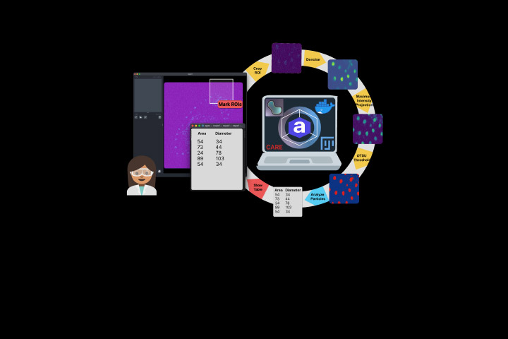
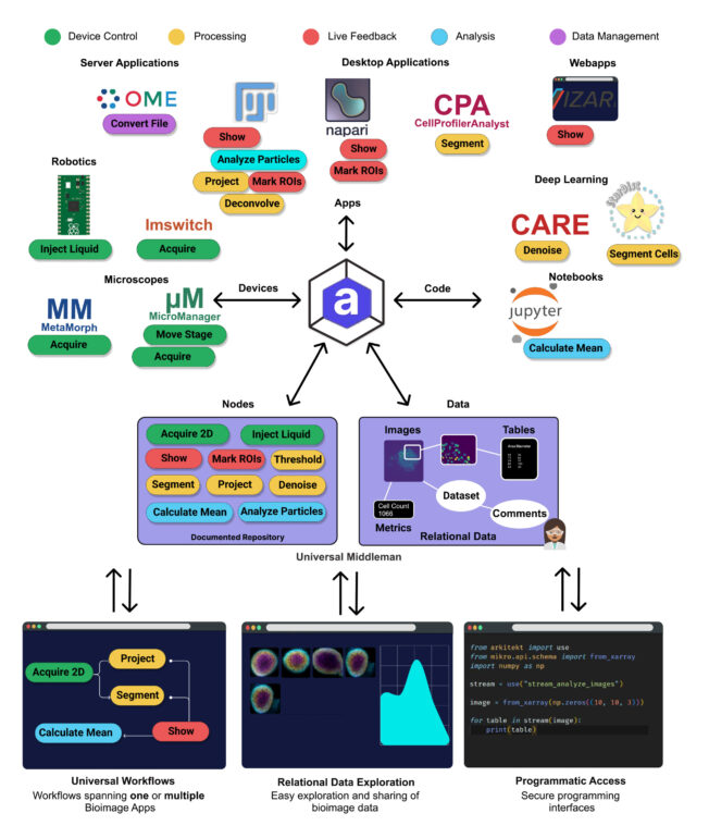
Arkitekt is an open-source software designed to facilitate microscopy image analysis tasks. It enables the creation of automated workflows for image processing and analysis, by intuitively orchestrating various bioimaging software on one or multiple computers, reliably and efficiently. Arkitekt integrates the most popular visualization and analysis software in bioimaging, such as Napari or ImageJ/FiJi, and can also execute code and control acquisition software.
Abstract
Quantitative microscopy workflows have evolved dramatically over the last years, progressively becoming more complex with the emergence of deep learning. Long-standing challenges like 3D segmentation of complex microscopy data can finally be addressed, and new imaging modalities are breaking records in both resolution and acquisition speed, generating gigabytes if not terabytes of data per day. With this shift in bioimage workflows comes an increasing need for efficient orchestration and data management, necessitating multi-tool interoperability and the ability to span dedicated computing resources. However, existing solutions are still limited in their flexibility and scalability and are usually restricted to off-line analysis. Here, we introduce Arkitekt, an open-source middleman between users and bioimage apps that enables complex quantitative microscopy workflows in real time. It allows orchestrating popular bioimage software locally or remotely in a reliable and efficient manner. It includes visualization and analysis modules, but also mechanisms to execute source code and pilot acquisition software, making ‘smart microscopy’ a reality.
Reference
Arkitekt: streaming analysis and real-time workflows for microscopy
Johannes Roos1, Stéphane Bancelin1, Tom Delaire1, Alexander Wilhelmi3 Florian Levet1,2, Maren Engelhardt3,4, Virgile Viasnoff5, Rémi Galland1, U. Valentin Nägerl1, Jean-Baptiste Sibarita*1
1 Univ. Bordeaux, CNRS, Interdisciplinary Institute for Neuroscience, IINS, UMR 5297, F-330006 Bordeaux, France
2 Univ. Bordeaux, CNRS, INSERM, Bordeaux Imaging Center, BIC, UAR3420, US 4, F-33000 Bordeaux, France
3 Frankfurt Institute for Advanced Studies (FIAS), 60438 Frankfurt am Main, Germany
4 Institute of Anatomy and Cell Biology, Medical Faculty, Johannes Kepler University, Linz, Austria
5 Mechanobiology Institute, National University of Singapore, Singapore
*Corresponding author:
Nature Methods ; 2024-09
https://doi.org/10.1038/s41592-024-02404-5
Mise à jour: 26/09/24

